

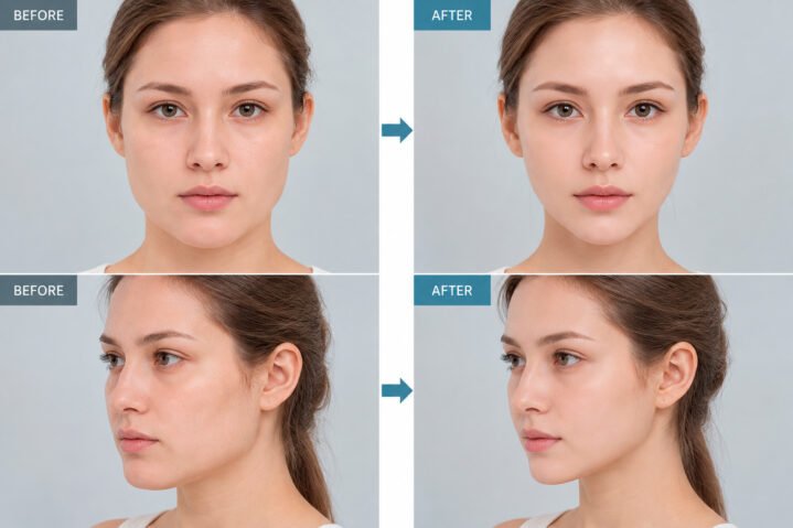
V-Line Surgery (Lower Jaw Contouring)
V-Line surgery is a facial contouring procedure designed to refine the shape of the lower jaw and create a slimmer, more balanced facial appearance. Depending on the patient's anatomy, the procedure may involve reshaping the mandibular angles and chin. As with any bone-contouring surgery, swelling and recovery can take several weeks, and the final results develop gradually. The goal is to achieve harmonious facial proportions while preserving the patient's natural features.
Who is this procedure for?
- You have a wide or prominent lower jaw
- You have a square or broad facial shape
- You have facial asymmetry related to the lower jaw
- You would like a slimmer, more defined jawline
Description of the procedure
The procedure is typically performed through incisions inside the mouth to avoid visible external scars. Depending on the surgical plan, the surgeon may reduce or reshape the mandibular angles and contour the lower jawbone. In some cases, outer cortical bone reduction may be performed to further narrow the lower face. The extent of bone reshaping is determined by the patient's facial anatomy, jaw structure, and desired outcome. A detailed examination and imaging studies are necessary to develop a safe and appropriate surgical plan.
Chin Reduction
Chin reduction surgery is designed to improve the proportions of a wide, long, or prominent chin. Depending on the patient's anatomy, techniques such as T-osteotomy or direct bone reduction may be recommended. In selected cases, the position of the chin can also be adjusted to improve facial balance and profile harmony. The surgical approach is tailored to the patient's individual facial structure and aesthetic goals.
Who is this procedure for?
- You have a wide or broad chin
- You have a prominent chin or concerns about chin projection
- You have mild chin asymmetry
- You have an elongated chin
- You would like to improve the proportions and contour of your chin
Description of the procedure
T-osteotomy
T-osteotomy is a chin-contouring technique that may be used to reduce chin width and adjust its shape. The surgeon typically makes an incision inside the lower lip, below the gum line, to access the chin bone. Carefully planned bone cuts allow the central portion of the chin to be reduced or repositioned. If necessary, additional bone reduction may be performed to shorten the chin. The remaining bone segments are then secured using fixation devices, such as titanium plates and screws. Resorbable fixation materials may be considered in selected cases. The bone edges are smoothed to create a more continuous and balanced chin contour. This procedure may also be combined with lower jaw contouring when appropriate.
Direct Resection (Bone Reduction)
Direct bone reduction may be suitable for patients who require a reduction in chin size or width without significant repositioning of the chin. The surgeon carefully removes or reshapes selected areas of bone to improve the chin contour. In certain cases, this technique may also help address minor asymmetry. The most appropriate method is determined following a detailed assessment of the patient's bone structure and facial proportions.
Cheekbone Reduction
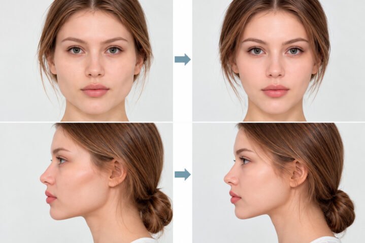
Cheekbone reduction, also known as reduction malarplasty or zygoma reduction, is a facial contouring procedure intended to reduce the prominence or width of the cheekbones. Prominent cheekbones can make the midface appear wider or more angular. By carefully repositioning the zygomatic bones, surgeons aim to create smoother facial contours and more balanced proportions while preserving the patient's natural appearance. The suitability of this procedure depends on the patient's bone structure, soft tissues, and individual aesthetic goals.
Who is this procedure for?
- You have a wide or prominent midface
- You have laterally protruding cheekbones
- You have prominent cheekbones that create an angular facial appearance
Description of the procedure
Cheekbone reduction is commonly performed through incisions inside the upper mouth, sometimes combined with small incisions near the hairline. The surgeon makes carefully planned bone cuts to reposition the cheekbone and zygomatic arch inward, reducing their prominence or lateral projection. The repositioned bone segments are typically secured with titanium plates and screws to support stability during healing. Resorbable fixation materials may be considered in selected cases. The surgical technique and extent of repositioning depend on the patient's facial anatomy, bone structure, and treatment goals. A thorough consultation and imaging assessment are essential before surgery.


Double Jaw Surgery (Orthognathic Surgery)
Double jaw surgery, also known as bimaxillary orthognathic surgery, is a procedure that repositions the upper and lower jaws to improve jaw alignment, facial proportions, and, when indicated, the patient's bite. It may be recommended for patients with significant skeletal discrepancies, facial asymmetry, or functional problems related to jaw position. The surgical plan is individually developed to achieve balanced facial contours while maintaining proper jaw function.
Who is this procedure for?
- You have facial asymmetry caused by differences in jaw position or development
- You have a protruding upper or lower jaw
- You have an excessively prominent lower jaw or an underbite
- You have a recessed or underdeveloped jaw
- You have an elongated lower face or significant vertical jaw imbalance
How is the procedure performed?
The surgical approach depends on the patient's skeletal structure, dental alignment, bite, and functional requirements. Before surgery, specialists typically perform a comprehensive examination, including dental assessments, X-rays, and 3D CT imaging when indicated. Digital surgical planning may be used to determine the appropriate repositioning of the jaws. Orthodontic treatment may be required before and/or after surgery, depending on the individual treatment plan.
Traditional Approach (Orthodontics Before Surgery)
In the traditional approach, orthodontic treatment is performed before jaw surgery to prepare the teeth for their planned positions after skeletal correction. This stage may take approximately 12–24 months, although the duration varies considerably. Once the teeth have been adequately prepared, surgery is performed to reposition the jaws. Additional orthodontic treatment is usually required after surgery to refine the bite and dental alignment.
Surgery-First Approach
The surgery-first approach allows selected patients to undergo jaw surgery before extensive orthodontic treatment. This method may provide an earlier improvement in facial appearance and reduce the overall treatment duration in suitable cases. Orthodontic treatment is subsequently performed to achieve proper dental alignment and bite stability. However, not every patient is a candidate for this approach. Eligibility depends on the existing bite, dental alignment, skeletal discrepancy, and detailed surgical and orthodontic planning.
Modified Surgery-First Approach
The modified surgery-first approach combines a shorter period of preoperative orthodontic preparation with jaw surgery and subsequent orthodontic treatment. In selected cases, preliminary orthodontic treatment may take approximately 3–4 months, although the duration depends on the patient's dental condition. This approach may be considered when limited tooth alignment is necessary before surgical correction.
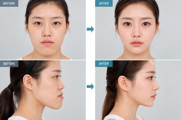
Other Jaw Surgeries
Upper Jaw Surgery (Le Fort I Osteotomy)
Le Fort I osteotomy is a commonly performed procedure for repositioning the upper jaw (maxilla). It may be used to correct skeletal discrepancies, improve dental occlusion, and address certain facial proportion concerns. The operation is performed under general anesthesia, usually through incisions inside the mouth to avoid visible external scars.
The surgeon carefully separates the upper jawbone using specialized instruments and repositions it according to the preoperative surgical plan. Depending on the patient's needs, the upper jaw may be moved forward, backward, upward, or downward. In selected cases, bone grafting may be necessary to support the planned repositioning.
Once the upper jaw is positioned correctly, it is typically secured with titanium plates and screws. The incisions are closed with sutures, which may be absorbable. Bone healing and recovery occur gradually over the following weeks and months. The final treatment plan depends on the patient's anatomy, bite, and functional requirements.
Lower Jaw Surgery
1) Sagittal Split Ramus Osteotomy (SSRO)
Sagittal split ramus osteotomy (SSRO), also known as bilateral sagittal split osteotomy (BSSO) when performed on both sides, is a surgical technique used to reposition the lower jaw. It may be recommended for patients with mandibular retrusion, mandibular prognathism, or certain bite abnormalities associated with skeletal jaw discrepancies.
The surgeon makes incisions inside the mouth and carefully divides the mandibular ramus on each side using planned bone cuts. This allows the tooth-bearing portion of the lower jaw to be moved forward or backward according to the surgical plan. The repositioned bone segments are typically stabilized with titanium plates and screws. Particular care is taken to protect surrounding nerves and anatomical structures, although temporary or persistent changes in sensation remain possible complications.
2) Vertical Ramus Osteotomy (VRO)
Vertical ramus osteotomy (VRO) is a lower jaw procedure that involves making a vertical bone cut through the mandibular ramus. An intraoral variation, known as intraoral vertical ramus osteotomy (IVRO), may be used to reposition the lower jaw, particularly in selected cases of mandibular prognathism or facial asymmetry.
The procedure allows the tooth-bearing portion of the mandible to be repositioned relative to the posterior segments. Depending on the technique, postoperative jaw stabilization and a period of restricted jaw movement may be required. VRO is generally used for selected mandibular setbacks rather than significant jaw advancement. The choice between VRO and SSRO depends on the patient's anatomy, bite, and surgical requirements.
3) Anterior Segmental Osteotomy (ASO)
Anterior segmental osteotomy (ASO) is a procedure used in selected patients with dentoalveolar protrusion, where the front teeth and supporting bone project excessively forward. Depending on the treatment plan, the procedure may involve the upper jaw, lower jaw, or both.
The procedure may require the removal of selected premolar teeth to create sufficient space for repositioning the anterior tooth-bearing bone segment. The segment is then carefully moved backward and stabilized using appropriate fixation devices. ASO may be considered for certain cases of protruding mouth appearance, but it is not suitable for every type of skeletal jaw discrepancy. A detailed orthodontic and surgical assessment is essential.
Recessed Chin Correction
A recessed chin is characterized by insufficient forward projection of the chin relative to the surrounding facial structures. It may result from the natural shape of the chin bone or from an underdeveloped or retruded lower jaw. These conditions require different treatment approaches. Depending on the patient's anatomy and bite, correction may involve chin augmentation, sliding genioplasty, or orthognathic surgery.
Who is this procedure for?
- You have a small or recessed chin
- You have an underdeveloped or retruded lower jaw
- You have bite problems associated with an abnormal jaw position
- You would like to improve chin projection and facial profile balance
Description of the procedure
a. Chin Augmentation with an Implant
Chin augmentation with an implant may be suitable for patients who have a small or recessed chin but otherwise normal jaw alignment and bite. The procedure is intended to increase chin projection and improve facial proportions.
The surgeon creates a small incision either inside the mouth or beneath the chin, depending on the selected technique. An appropriately sized implant, commonly made of medical-grade silicone or another suitable material, is positioned over the chin bone. The procedure may be performed under local anesthesia with sedation or general anesthesia, depending on the patient's needs and the surgeon's recommendation. Recovery time varies, and swelling or temporary discomfort may occur.
b. Sliding Genioplasty (Chin Advancement)
Sliding genioplasty is a bone-repositioning procedure used to correct chin projection without necessarily changing the position of the entire lower jaw. It may be suitable for patients whose bite is acceptable but whose chin is recessed or disproportionate to the rest of the face.
The surgeon makes an incision inside the lower lip to access the chin bone. A carefully planned bone cut allows the lower portion of the chin to be moved forward. Depending on the patient's needs, the chin may also be repositioned vertically or adjusted to improve symmetry. The repositioned bone segment is typically secured with titanium plates and screws. The procedure is planned with consideration for nearby tooth roots and sensory nerves.
c. Lower Jaw Advancement (Mandibular Advancement Surgery)
Lower jaw advancement surgery may be recommended when a recessed chin is associated with an underdeveloped or retruded mandible and an abnormal bite. Unlike isolated chin augmentation, this procedure repositions the lower jaw, including the tooth-bearing segment, to improve skeletal alignment and occlusion.
The procedure is commonly performed using bilateral sagittal split osteotomy (BSSO). Through incisions inside the mouth, the surgeon carefully divides the lower jawbone and moves the mandible forward according to the preoperative plan. The repositioned bone segments are typically secured with titanium plates and screws. Orthodontic treatment may be required before or after surgery. In certain cases, surgery on both jaws may be necessary to achieve appropriate functional and skeletal alignment. Recovery, dietary restrictions, and return to normal activities vary depending on the extent of surgery and individual healing.
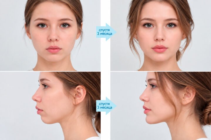
Forehead Contouring and Augmentation
Forehead augmentation is an aesthetic procedure designed to improve the contour and projection of a flat, recessed, or underdeveloped forehead. Depending on the patient's anatomy and treatment goals, the procedure may involve a customized implant or autologous fat grafting. The objective is to create smoother forehead contours and balanced facial proportions while preserving a natural appearance.
Who is this procedure for?
- You have a flat forehead
- You have a recessed or underprojected forehead
- You would like a smoother or more rounded forehead contour
Description of the procedure
a. Customized Implant Preparation
For patients undergoing forehead augmentation with an implant, the surgeon may recommend a customized implant based on the patient's forehead anatomy. Imaging studies and digital planning may be used to determine the appropriate implant shape, thickness, and position. The implant is prepared and assessed before surgery to support a balanced forehead contour.
b. Forehead Implant Surgery
Forehead implant placement is typically performed through an incision within the scalp or near the hairline, depending on the surgical approach. The surgeon creates a suitable space and positions the implant to improve forehead projection and contour. The procedure may be performed under general anesthesia or another appropriate form of anesthesia, depending on the surgical plan. Temporary swelling, discomfort, and changes in sensation may occur during recovery.
c. Forehead Fat Grafting (Autologous Fat Transfer)
Forehead fat grafting is a procedure that uses the patient's own fat to improve forehead volume and contour. Fat is typically harvested from areas such as the abdomen or thighs through liposuction, then processed and carefully injected into selected areas of the forehead using small entry points. The goal is to achieve smooth volume distribution and natural-looking contours. Some of the transferred fat may be reabsorbed during healing, and additional treatment may be necessary to achieve or maintain the desired volume.
Paranasal Augmentation
Paranasal augmentation is an aesthetic procedure designed to improve volume and projection in the areas beside the base of the nose. It may be considered for patients with a recessed paranasal region, which can influence the appearance of the midface and nasolabial folds. Depending on the patient's anatomy, augmentation may be performed using implants or other suitable techniques.
Who is this procedure for?
You have a recessed or underprojected area beside the nose and would like to improve midfacial balance and facial contours.
Description of the procedure
Paranasal implant augmentation is commonly performed through small incisions inside the upper mouth. The surgeon carefully creates space over the bone near the base of the nose and positions an appropriately shaped implant to improve projection in the paranasal region. Implant materials may include medical-grade silicone or other biocompatible materials selected according to the surgical plan. The size, shape, and position of the implant are determined based on the patient's facial anatomy and aesthetic goals. As with other implant procedures, possible complications include infection, implant displacement, asymmetry, and the need for revision surgery.
#
R
E
A
L
S
T
O
R
Y

GG Story



#RealStory